*** Advanced Project Notice ***
The following project is an ADVANCED project and may require handling of dangerous materials and/or equipment and is intended to be conducted by adults only!

Purpose
To isolate DNA from yeast (a unicellular fungus).
Additional information
Isolation of DNA from living cells is very important in molecular biological studies. You may have heard about DNA finger printing, genetic engineering…etc; all these molecular biological works in need of isolated pure DNA. Isolation of chromosomal DNA or plasmid DNA of prokaryotic organisms in pure state is not an easy target. It has to be carried out with great care. Most of the students are interested in DNA and wonder how to isolate DNA from living entities.
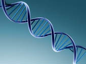
In higher plants, higher animals and in most other cellular organisms, genetic information are stored as base sequences in DNA molecules. These are very large molecules with very high molecular masses. DNA consists of a double helix. The two strands in one molecule are complementary and antiparallel to each other. Hydrogen bonds between nitrogenous bases of two strands keep them together. Inside the cell, DNA exist as DNA-protein complexes. The associated protein is called histamine.
Different compounds absorb visible light or UV radiation of different wave lengths. Therefore the wave length of light absorbed by a certain compound is unique. This special property can be used in identification of chemical compounds. DNA absorb strongly in the UV region due to the conjugated double bond system of constituents purine and pyrimidines (nitrogenous bases). It shows characteristic maximum absorbance at 260nm and a minimum at 230nm. A special instrument called colorimeter is used to measure absorbance values.
Sponsored Links
Required materials
- Graduated cylinder(10ml)
- Erlenmeyer flasks (100ml)
- Centrifuger
- 15 ml Test tubes
- A glass rod
- 0.5g of Lysozyme
- 5M NaCl solution
- Saline- EDTA solution:- prepare to contain 0.15M NaCl and 0.1M EDTA
[Preparation of 1.0M EDTA solution: - 292.24g of disodium ethylene diamine tetra acetate.2H2O in1000ml distilled water. Adjust the pH to 8 with NaOH (~20g). Autoclave]
- 25% (W/V) sodium dodecyl sulfate (SDS)- a detergent
- 25 ml detergent
- 100 ml distilled water
- TE buffer(1mM Tris, 1mM EDTA, pH8)
Molecular weight of Tris base = 121.14 g mol-1/ Molecular weight of EDTA = 292.24 g mol-1- 0.121g Tris
- 0.292g EDTA
- 1000 ml distilled water
- 2g of Bakers yeast
- Chloroform : Isoamyl alcohol (24:1) solution
- 95% ice cold ethanol
Estimated Experiment Time
3 to 4 hours
Step-By-Step Procedure
- 1. Take an Erlenmeyer flask. Suspend 2g of pre weighed baker’s yeast sample in 6ml of saline- EDTA solution. Heat up to 50ºC in a hot water bath to dissolve yeast properly.
- 2. Add 0.5g of Lysozyme and incubate at 37ºC for 30 min.
- 3. Add 1.0ml of 25% SDS and heat in a water bath (60ºC) for 10 min. swirl the flask slowly (solution will become viscous due to release of DNA).
- 4. Cool to room temperature, add 4.5 ml of 5M NaCl and mix well.
- 5. Add equal volume (~12ml) of chloroform : isoamyl alcohol solution and mix thoroughly. Pour the suspension into 15ml test tubes and centrifuge the suspension at 8000rpm for 5 min.(3 layers must be visible, upper aqueous phase with nucleic acids, middle interface with denatured proteins and bottom organic phase with lipids and cell debris.)
- 6. Carefully transfer the upper layer with a dropper into a 100 ml Erlenmeyer flask or to a large test tube.
- 7. Add two volumes (as twice as the volume of decant aqueous layer) of ice cold 95% ethanol. Must be poured down by the side of the container. Swirl for 10 min at room temperature. In this step you will see DNA precipitation. Alternatively the precipitation can be done at -20ºC for 20 min.
- 8. Spool the DNA into glass rod. If DNA does not form fibrous threads upon addition of ethanol, centrifuge the mixture at 4000rpm and wash the DNA pellet (sediment) gently with 70% ethanol (pour EtOH slowly by the wall to wet the pellet. Then decant slowly without disturbing the pellet). Allowed to dry and store at -80ºC as a dry pellet.
- 9. To determine that the isolated pellet actually contains DNA, dissolve the pellet in sterile TE buffer and measure the absorbance in a range of wavelengths (from about 200nm to 300nm). Plot a graph absorbance vs. wave length. If you get a maximum absorbance at 260nm your prepared solution contains DNA. If the solution is not pure DNA but a mixture of DNA and protein impurities then you will see a high absorbance at 280 nm too.
Note
*** THIS IS AN ADVANCED SCIENCE PROJECT ***
Observation
Chromosomal DNA is thin and long. It is better to avoid excessive agitation as much as possible, during the isolation process. Here we add a number of solutions to isolate DNA as pure as possible. You might wonder why we add so many solutions. Following list contains the purpose of each solution we add.
- Lysozyme – degrade the polysaccharides in yeast cell wall. By breaching the cell wall this enzyme facilitates DNA movement into external liquid.
- The release DNA is buffered in saline-EDTA
- EDTA serves as a chelating enzyme and removes Mg2+ ions from the solution. Without Mg ions DNAse enzymes released by the cell can not degrade DNA. So we can isolate DNA as long strands. Citrate buffer can also be used to achieve same aspect.
- pH is maintained in 8 to reduce the interaction of histamine proteins with DNA.
- SDS detergent denatures proteins. Hence DNA-protein complexes are dissociated.
- High salt buffer (5M NaCl) – diminish the ionic character of DNA causes complete dissociation of DNA-protein complex.
- Chloroform : isoamyl alcohol addition followed by centrifugation – dissociated proteins are removed from DNA extract.
Result
At the eighth step, if you put a glass rod into the DNA extraction thin strands of DNA will spool onto the glass surface. If some how you couldn’t see this phenomenon due to excessive break down of chromosomal DNA into small pieces; measure the absorbance of DNA solution in UV range. If you have isolated pure DNA, the maximum absorbance will turn out at 260nm. By examining the plot you prepare as described in step 9, you will be able to get an idea about the purity of DNA.
Sponsored Links
Take a moment to visit our table of Periodic Elements page where you can get an in-depth view of all the elements,
complete with the industry first side-by-side element comparisons!